Section outline
-
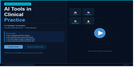
Welcome to Week 3! This module shifts into applied clinical practice—examining how AI tools are utilized day-to-day within live medical imaging environments. Through realistic case scenarios, learners will develop the practical judgment required to work effectively alongside clinical AI software, understanding both when to trust automated alerts and when to question them.
🔑 Key Operational Topics Covered:
-
System Integration: Real-world AI workflow integration covering triage systems, worklist prioritization matrixes and direct notification alerts.
-
Professional Protocols: The Radiologic Technologist's (RT) precise role when AI flags an image—focusing on documentation standardizations, escalation paths and communication responsibilities.
-
Quality Assurance (QA): Technical quality control considerations including anatomy positioning errors, patient motion artifacts and how they directly alter AI performance outputs.
-
System Vulnerabilities: Understanding AI "blind spots"—exploring exactly what current algorithmic models miss and why.
-
Clinical Case Reviews: Real-world radiology department case studies focusing on AI success stories and near-miss diagnostics prevention.
🎯 Week 3 Learning Outcomes:
By the end of this week, you will be able to:
-
Demonstrate a clear understanding of how AI triage and worklist prioritization tools interact dynamically with existing RT imaging workflows.
-
Articulate your professional responsibilities and clinical escalation limits when an automated system flags an urgent pathology.
-
Identify at least three distinct image quality degradation factors (e.g., patient motion, standard artifacting, bad positioning) that cause AI interpretation failures.
-
Apply communication best practices when relaying critical AI-flagged findings to supervising radiologists safely.
-
Students mustViewComplete interactions 100%Watch till the endReceive a gradeReceive a passing gradeAI Tools in Clinical Practice — Week 3 Interactive Video0% (0/1) 0/20
-
Students mustViewFinish all steps in the activityReceive a gradeReceive a passing grade
-
Students mustViewMake forum posts: 1
After completing the interactive walkthrough on AI Triage Dashboards, share your perspective with your peers.
Have you encountered AI tools or automated alerts in your clinical department? How do you think these systems—like the PACS integration we reviewed—alter your daily imaging workflow or decision-making process?
-